|
In japan, deep
mycosis, an opportunistic infectious disease,tends to increase yearly.
Its clinical diagnosis is difficult, in many cases, and depends
on pathologic histology. The Limulus reagent(LAL : Limulus amebocyte
lysate), made from extract of blood cells of horses- |
|
|
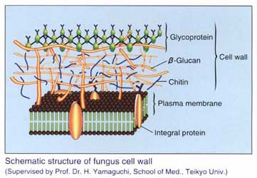
-β-D-glucanase. Various types of β-(1→6) branched (1→3)-β-D-glucans
were isolated from fungi, with biological activities such as antitumor
activity. In case of deep mycosis, infecting fungi release (1→3)-β-D-glucan
in the blood stream. Diagnosis of deep mycosis utilizing 1→3)-β-D-glucan
as a diagnosing parameter has been explored. Wako β-Glucan Test
measures (1→3)-β-D-glucan by the kinetic turbidimetric assay in
a sample pretreated with a solution which inactivats endotoxin in
the sample by use of non-ionic detergent and polymyxin B. |
|
|
|
|
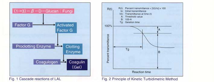
Endotoxin in a sample is inactivated by treating the sample at 80℃ for 10 miunutes with the pretreatment solution, which contains non-ionic detergent and polymyxin B. This pretreatment also inactivates inhibitory protein substances in the sample. When the pretreated sample is mixed with the LAL solution, (1→3)-β-D-glucan in the sample activates Factor G, which initiates the cascade reactions shown in Figure 1, and causes gelation. The turbidity change caused by the gelation reaction is detected as transmittance change. The time taken for the transmittance to reach the threshold value is also measured. This time is defined as gelation time(Tg). (Figure 2) The Log(β-glucan concentration) is in inverse proportion to log[log(Tg)]. Wghen the Tg of an unknown sample is measured, the β-glucan concentration of the sample can be obtained from a standard curve. (Kinetic Turbidimetric Method) |
|
|