Caspase™ Apoptosis Fluorometric Assays
본문
|
Caspase™ Apoptosis Fluorometric Assays
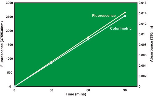
The CasPASE™ Apoptosis assays are designed to monitor apoptosis by measuring various caspase (protease) activities, a key early indicator of apoptosis in mammalian cells. The assay provides a simple and easy to follow method that can be monitored with a fluorescence reader. The assay is based on the detection of cleavage of a synthetic substrate, which has 7-amino-4- trifluromethyl coumarin (AFC) at the C-terminal. When liberated from the peptide, AFC produces an optical change that exhibits a fluorescence spectral shift between the substrate-conjugate and the free dye. The AFC substrate is fluorogenic (detected at 510 to 550 nm with a fluorometer). During the reaction, the substrate releases AFC free dye and undergoes a fluorescence shift. The reaction is selectively and irreversibly inhibited by the peptide Z-VAD-FMK (fluoromethyl ketone). Comparison of the fluorescence from an induced/apoptotic sample with an uninduced control allows one to determine the fold-increase in protease activity. The Caspase activity can also be quantitated by using a standard curve established with an appropriate free dye. When measuring fluorescence, the assay reaction is excited at 380 to 400nm and emission is read at 510 to 550nm. The assays can be conveniently adapted for high-throughput 96-well format. The assay system may be used with purified enzyme preparations, cell extracts or tissue lysates. CasPASE™ assays are available for caspase enzymes 1 through 10 and 13. Each CasPASE™ assay kit is supplied with necessary assay buffers, enzyme specific AFC-substrate, free dye (AFC), and the potent caspase inhibitor Z-VAD-FMK for establishing proper positive and negative controls and standards.
Colorimetric Caspase Assays are also available.
Features • Assays for caspase enzymes 1 through 10 and 13 are available • Exhibit fluorescence spectral shift • Conveniently adaptable for high-throughout 96-well format • All kits supplied with enzyme specific AFC-substrate, free dye (AFC) & inhibitor Z-VAD-FMK
Applications • Monitor apotosis by measuring various caspase activities in cells and tissues • Use with purified enzyme preparations, cell extracts, and tissue lysates • Characterization of caspase enzymes
References
Instruction sheet (pdf)
|
Ordering informations
|
Catalog No. |
Product Name |
Size |
|
786-200A |
Caspase™ 1, 4, 5 assay with Ac-WEHD-AFC substrate |
50 assays |
|
786-200B |
Caspase™ 1, 4, 5 assay with Ac-WEHD-AFC substrate |
100 assays |
|
786-201A |
Caspase™ 2 assay with Ac-VDVAD-AFC substrate |
50 assays |
|
786-201B |
Caspase™ 2 assay with Ac-VDVAD-AFC substrate |
100 assays |
|
786-202A |
Caspase™ 3, 7, 10 assay with Ac-DEVD-AFC substrate |
50 assays |
|
786-202B |
Caspase™ 3, 7, 10 assay with Ac-DEVD-AFC substrate |
100 assays |
|
786-203A |
Caspase™ 6 assay with Ac-VEID-AFC substrate |
50 assays |
|
786-203B |
Caspase™ 6 assay with Ac-VEID-AFC substrate |
100 assays |
|
786-204A |
Caspase™ 8 assay with Ac-LETD-AFC substrate |
50 assays |
|
786-204B |
Caspase™ 8 assay with Ac-LETD-AFC substrate |
100 assays |
|
786-205A |
Caspase™ 9 assay with Ac-LEHD-AFC substrate |
50 assays |
|
786-205B |
Caspase™ 9 assay with Ac-LEHD-AFC substrate |
100 assays |
|
786-206A |
Caspase™ 13 assay with Ac-LEED-AFC substrate |
50 assays |
|
786-206B |
Caspase™ 13 assay with Ac-LEED-AFC substrate |
100 assays |
▣ 관련 페이지 ; G-Biosciences
댓글목록
등록된 댓글이 없습니다.