Probe, target & illuminate only the Cancer Cell Mitochondria
본문
|
AIE Cancer Yellow, Target and illuminate only the Cancer Cell Mitochondria 생세포와 고정세포 모두에 사용 할 수 있는 암세포 특이적 형광 프로브
일반적인 형광물질은 ACQ(Aggregation Caused Quenching) 방식으로 응집하면서 발광이 약해지는 문제가 있습니 다. AIEGEN Biotech 사의 probe는 AIE(Aggregation-Induced Emission) 방식으로, 응집되면서 집합체를 형성하면 강한 형광을 발합니다.
The product can be used for quick cell imaging as well as fixed localized imaging. The product can serve as a photosensitizer to generate reactive oxygen species (ROS) to induce cell apoptosis, which can be used for photodynamic therapy. The product can be excited by 405 nm laser of confocal microscope after co-cultured with cell and the greenish-yellow signal can be collected above 500 nm.
Specifications; • Purpose: Mitochondria staining and induce cell apoptosis • Color: Orange • Imaging platform: Fluorescence microscope, Confocal microscope • Pack size and quantity: 10 μmol • Detection method: Fluorescence • Excitation / Emission (nm): 430±20 / 560±50 • Recommended transport condition: Room temperature • Product declaration: Only used for research. Do not apply to any detection procedure.
The product has been tested working on • Human cervix HeLa • Human breast MDA-MB-231 • Human lung HCC827 • Human breast MCF-7 • Human lung A549 • Human liver HepG2 • Human lung PC-9
Examples of use;
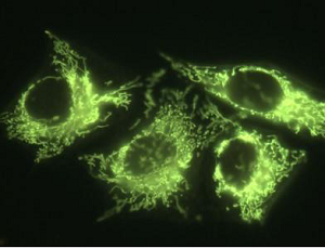
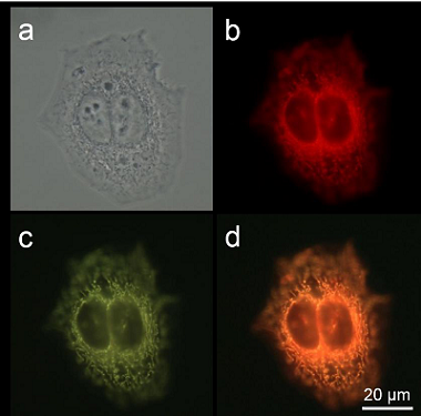
Figure 1: Co-localization imaging of HeLa cells stained with MitoTracker Red and AIETM Cancer Yellow. (a) Bright-field image and (b and c) fluorescent images of HeLa cells stained with (b) MTR (50 nM) and (c) AIETM Cancer Yellow (200 nM) for 20 min. (d) Merged image of panel (b) and (c). λex: 540~580 nm (MTR) and 400~440 nm (AIETM Cancer Yellow); scale bar = 20 μm.
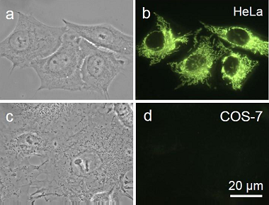
Figure 2: Differentiation of cancerous HeLa cells from normal COS-7 cells by AIETM Cancer Yellow. (a and c) Bright-field and (b and d) fluorescent images of (a and b) HeLa cells and (c and d) COS- 7 cells incubated with 200 nM of AIETM Cancer Yellow for 20 min.
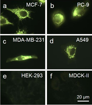
Figure 3: Differentiation of cancer cells from normal cells by AIETM Cancer Yellow. (af) Fluorescent images of different (ad) cancer cells and (ef) normal cells stained with AIETM Cancer Yellow (200 nM) for 20 min. λex: 400~440 nm.
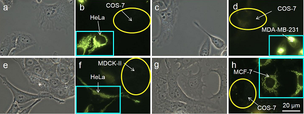
Figure 4: Co-culture different combinations of cancer cell and normal cell in culture medium with AIETM Cancer Yellow. (a, c, e and g) Bright-field images and (b, d, f and h) corresponding fluorescent images of (a and b) HeLa and COS-7 cells, (c and d) MDA-MB-231 and COS-7 cells, (e and f) HeLa and MDCK-II cells and (g and h) MCF-7 and COS-7 cells incubated in Dulbecco’s Modified Eagle Medium (DMEM) with AIETM Cancer Yellow. Light blue rectangles represent cancer cells and yellow circles represent normal cells. All the images share the same scale bar: 20 μm.
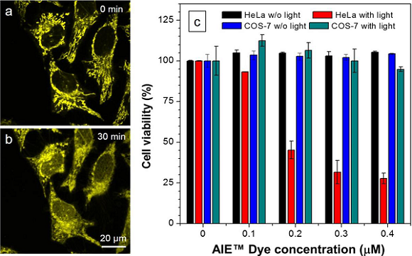
Figure 5: AIETM Cancer Yellow selectively kills cancer cells through PDT. (a,b) Change in mitochondrial morphology before and after white light irradiation. (c) Cell viability of HeLa cells and COS-7 cells stained with different concentrations of AIETM Cancer Yellow in the absence or presence of white light irradiation. |
Ordering information;
|
Catalog No. |
Product Name |
Size |
|
N/A |
AIETM Cancer Yellow |
10 μmol |
▣ 관련 페이지 ; AIEGEN Biotech
댓글목록
등록된 댓글이 없습니다.